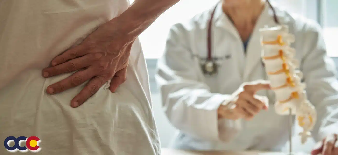
Thoracic fractures can be extremely painful, particularly if there is displacement of bone fragments or injury to surrounding soft tissues. Delayed treatment of thoracic fractures can increase the risk of complications such as spinal instability, deformity, nerve damage, and paralysis. All of which may be permanent and irreversible. In the case of thoracic fracture, you need a surgeon at the top of their field. Someone whose experience and skill you can trust. At OCC – Advanced Orthopedics & Sports Medicine Specialists in Denver, Parker, or Aurora, Colorado, their highly accomplished surgeons are known and respected for their diagnosis and treatment of thoracic fractures. When it comes to thoracic fracture, you can count on getting not just the best treatment but the best results.
OVERVIEW
People sometimes refer to a spinal fracture as a broken back. When a bone in the spine collapses, it is called a vertebral compression fracture (VCF). Vertebral compression fractures are the most common injury to the thoracic spine. Osteoporosis causes more than 1.5 million vertebral compression fractures each year. They are almost twice as common as other fractures typically linked to osteoporosis, such as broken hips and wrists. Forty percent of all women will have at least one vertebral compression fracture by the time they are 80 years old. More than 150,000 thoracic fractures each year are caused by traumas. Men are 4 times more likely to have a traumatic spinal fracture than women. Once someone has had a vertebral compression fracture, they are five times more likely to develop another compared to someone who’s never experienced one. Prognosis and recovery from a thoracic fracture may differ from patient to patient. The difference is due to the type of injury and the level of severity.
ABOUT THE THORACIC SPINE
The spine is made up of three segments. When viewed from the side, these segments form three natural curves. The “c-shaped” curves of the neck (cervical spine) and lower back (lumbar spine) and the “reverse c-shaped” curve of the chest (thoracic spine). 33 vertebrae make up the spine. Vertebrae are the individual bones that interlock with each other to form the spinal column. The thoracic spine consists of 12 vertebrae numbered T1 to T12. Each number corresponds with the nerves in that section of the spinal cord. These nerves branch off, transmitting signals between the brain and major organs, including the lungs, heart, liver, and small intestine. Thoracic vertebrae are unique in that they have the additional role of providing attachments for the ribs. This makes the thoracic spine more rigid and stable than the cervical or lumbar regions, making it the least common area of injury along the spine. While it’s primarily designed for stability, force absorption, and keeping the body upright, the thoracic spine is capable of a wide range of movement and its mobility is vital to overall health and function.
WHAT IS THORACIC FRACTURE?
There are several types of thoracic fractures, which can vary based on the mechanism of injury, severity, and location within the thoracic spine. These include compression fractures (wedge), burst fractures, flexion-distraction (seat belt injury/Chance fracture), and fracture-dislocation. A stable versus an unstable fracture is another way a provider will classify a thoracic fracture. With a stable fracture, the injury that broke the vertebrae didn’t push or pull them out of their usual place in the spine. One is less likely to need surgery with a stable fracture. Unstable fractures happen when the injury moves the vertebrae out of their usual alignment. There’s a much higher chance of the need for surgery to repair the broken vertebrae and a higher risk for dangerous complications that can affect the spinal cord.
Read more about Thoracic Fractures on our new Orthopedic News Site – Colorado Orthopedic News. Schedule an appointment with a spine specialist today.


